
Fig 6. In vivo sub-surface vascular imaging with ICG and IR-E1050.
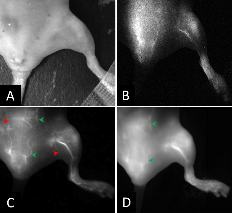
Representative images of the hind limb in a mouse in; (A) visible light, (B) NIR-II window obtained after i.v. administration of IR-E1050, (C) NIR-II window obtained after i.v. administration of ICG, and (D) NIR-I window after i.v. administration of ICG. (C) and (D) is the same animal. NIR images at 5 minutes post injection.