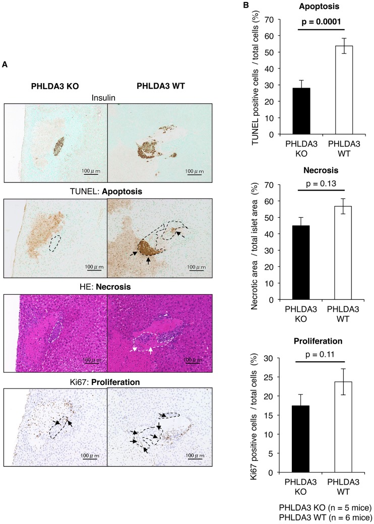
Fig 4. Condition of islets at 12 hs after transplantation.
A. Histological analyses of KO and WT islets at 12 hs after transplantation. Apoptosis (TUNEL-positive), necrosis (eosin-positive without nucleus area) and proliferation (Ki67-positive) were seen in both the KO (n = 5) and WT (n = 6) groups, but they were less prominent in the KO group. The images (insulin, TUNEL, HE, Ki67) were of the same location. The dashed lines indicate the border of the transplanted islets. The size of the scale bar was 100 μm. B. Quantification of the digitalized images showing apoptosis, necrosis and proliferation. The data confirms that apoptosis of transplanted islets at 12 hs was lower in the KO group. The statistical analysis was done using the Mann–Whitney U test.

