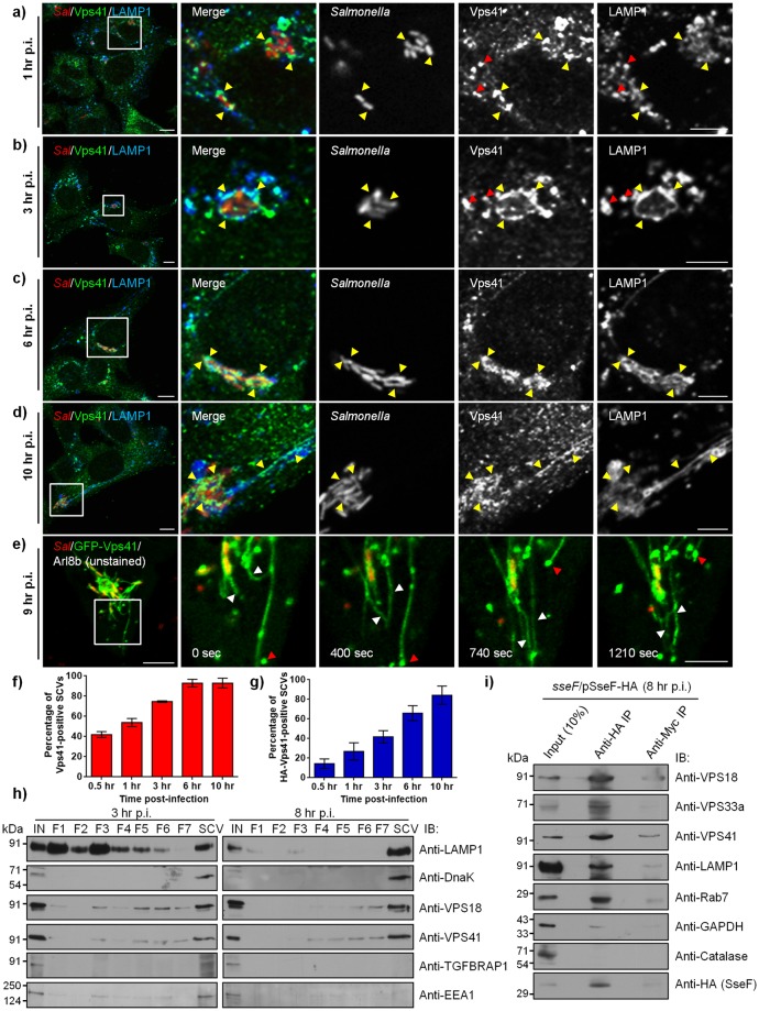
Fig 1. HOPS subunits are recruited to LAMP1-positive SCVs and SIFs during Salmonella infection.
a-d) Representative confocal micrographs of HeLa cells infected with DsRed-expressing Salmonella (red). At different time points post infection (p.i.), cells were fixed and stained for endogenous Vps41 (green) and LAMP1 (blue). Different panels represent a higher magnification of the boxed areas, showing recruitment of Vps41 on SCVs and SIFs (marked by yellow arrowheads). Red arrowheads indicate lysosomal localization of Vps41 in panels (a) and (b). Bars: (main) 10 μm; (insets) 5 μm. e) Time-lapse microscopy of HeLa cells co-transfected with plasmids encoding GFP-Vps41 and untagged-Arl8b, and infected with DsRed-expressing Salmonella (red). Time-lapse series were recorded 9 hr p.i., and still images shown here correspond to S2 Movie. Different panels represent a higher magnification of the boxed area indicating Vps41-positive SIFs emanating from the SCVs showing extension, retraction and bifurcation (white arrowheads). Red arrowheads indicate fusion of Vps41-positive vesicles with SIFs. Bars: (main) 10 μm; (insets) 5 μm. f and g) Quantification of endogenous (f) or HA-tagged Vps41 (g)-positive SCVs at different time points p.i. Data represent percentage of Vps41-postive SCVs scored for ~100 SCVs for each time point. The mean ± S.D. is shown for three independent experiments. h) SCVs were isolated from Salmonella-infected HeLa cells at 3 hr and 8 hr p.i. using sucrose density ultracentrifugation, followed by second round of ultracentrifugation of fractions 8–10 on a ficoll cushion (labeled as SCV). Different fractions were resolved on SDS-PAGE gel and immunoblotted using indicated antibodies. i) Salmonella-modified membranes were isolated from HeLa cells infected with sseF-deficient strain of Salmonella harboring an expression vector with a C-terminal epitope-tagged sseF and its cognate chaperone sscB (sseF/pSseF-HA) at 8 hr p.i. by differential centrifugation. The enriched fraction was further subjected to affinity immunoprecipitation (IP) using anti-HA antibody-conjugated agarose beads or anti-Myc antibody-conjugated agarose beads as a control. The eluted samples were analyzed for presence of effector protein (SseF) and host proteins by Western blotting as indicated.

