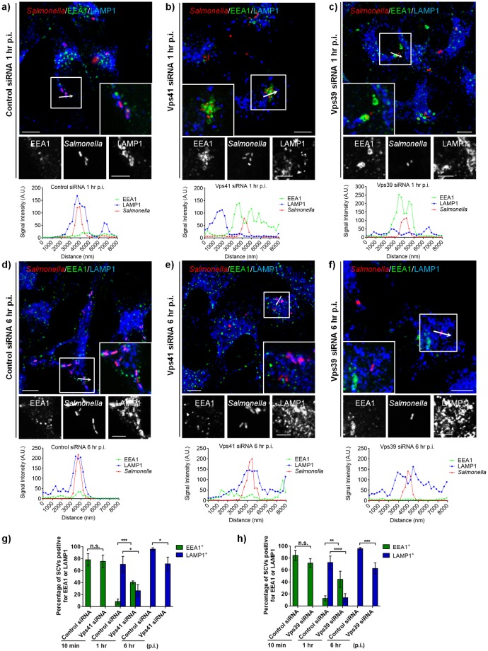
Fig 3. Depletion of HOPS subunits delays but does not block SCV maturation.
a-f) Representative confocal micrographs of control siRNA-, Vps41 siRNA- or Vps39 siRNA-treated HeLa cells infected with DsRed-expressing Salmonella (red). At different time points p.i., cells were fixed and stained for early endosomes marker, EEA1 (green), and LAMP1 (blue). Insets depict higher magnification of the boxed areas showing localization of different markers on the SCVs. Intensity line scan profile of EEA1/LAMP1 across the width of a single SCV (indicated by an arrow in the boxed region) is shown below the individual image. Bars: (main) 10 μm; (insets) 5 μm. g and h) Quantification of percentage of infected cells displaying EEA1/LAMP1-accumulation around SCVs at the indicated time point p.i. Data represent mean ± S.D. for ~50 SCVs from three independent experiments (n.s., not significant; *, P < 0.05; **, P < 0.01; ***, P < 0.001; ****, P < 0.0001; Student’s t test).

