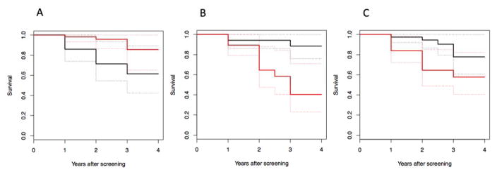
Figure 1.
Kaplan-Meier curves of a median split of imaging outcome measure. Black curves are for subjects with greater than the median and red curves are less than the median imaging signal. Dashed lines are the 95% confidence interval lines. A: Higher [11C]PIB binding in the precuneus is associated with greater rates of progression to dementia. B: Lower [18F]FDG SUVR in the parietal cortex is associated with greater rates of progression to dementia. C: Lower hippocampus volume is related to greater rates of progression to dementia.

