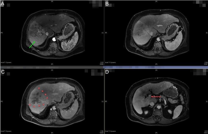
Figure 1. Pre-treatment diagnostic magnetic resonance imaging (MRI) of the abdominal scan.
Multi-phase liver protocol MRI abdomen axial images showing arterial (A), venous (B), and delayed (C) phases of the diffusely infiltrative and heterogeneously enhancing hepatocellular carcinoma tumor involving segments five through eight (best seen on panel C, arrowheads). The portal vein tumor thrombus is seen extending into the main portal vein on venous delay (D - red arrow). Peri-tumoral perfusion abnormalities are seen in panel A (green arrow).

