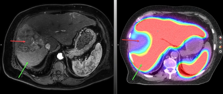
Figure 2. The treatment planning sulfur colloid single-photon emission computed tomography (SPECT/CT) scan.
The technetium-99m [99mTc] sulfur colloid SPECT/CT scan with which images sulfur colloid uptake in liver Kupffer cells and has been shown to correlate with global clinical liver function, was used to help identify and delineate both gross tumor and uninvolved normal liver. Areas of high sulfur colloid uptake represent non-GTV liver (red color wash) and the large well-defined photopenic defect (red arrow) corresponds to the tumor. The presumed perfusion abnormalities seen on the magnetic resonance imaging (MRI) scan (left panel) show high uptake of sulfur colloid on SPECT/CT (green arrows) and provides confirmation that these areas in the liver are unlikely infiltrated with tumor (red arrow).

