Figure 8.

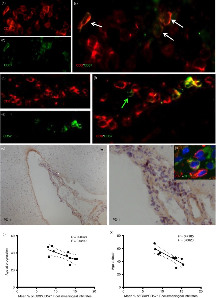
CD3+ CD57+ T cells are present in inflamed perivascular and meningeal infiltrates of post‐mortem secondary progressive multiple sclerosis (SP‐MS) brain tissue. Double immunofluorescence staining was performed on post‐mortem SP‐MS brains. CD3+ CD57+ cells (white arrows) were detected in meningeal infiltrates containing substantial numbers of CD3+ cells (a–c). Also, similar numbers of CD8+ CD57+ cells were detected in the same infiltrates (d–f). However, occasional CD8‐ CD57+ cells were also observed (green arrow, f). By using immunohistochemistry assessment of programmed death 1 (PD‐1) expression on serial sections from the same post‐mortem MS cases, several scattered PD‐1+ cells have been detected in particular in meningeal infiltrates (g and h, higher magnification of a selected area *). Double immunofluorescence demonstrated that a substantial proportion of the PD‐1+ cells infiltrating the meninges are CD57+ PD1+ cells (inset i in h). Negative correlation was found in the meningeal infiltrates between % of CD3+ CD57+ cells and age at disease progression (k) (r = 0·4648, P = 0·0299) and age at death (j) (r = 0·7185; P = 0·0020).
