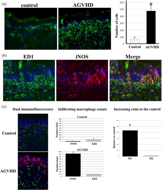
Figure 2.

Activated macrophages accumulate and persist in oral mucosal acute graft‐versus‐host disease (AGVHD). (a) Immunofluorescent images of ED1‐positive macrophages in the tongue from the control and AGVHD‐mediated rats. Original magnification, ×200. For the quantitative analysis of macrophage numbers, 10 areas 50 µm2 each in the tongue were selected randomly from each of the five rats and the total number of ED1‐positive cells was counted. The mean number of ED1‐positive cells per 10 areas ± standard deviation (s.d.) is shown for each group. *Significant difference at P < 0·01 (Student's t‐test). (b) Dual immunofluorescent images of ED1 (green) and inducible nitric oxide synthase (iNOS) (red) in oral mucosal AGVHD. The nucleus was stained with Hoechst 33324 (blue). Original magnification, ×200. (c) Distribution of M1 and M2 macrophages using iNOS (red) and ED2 (green) antibodies, respectively. Original magnification, ×200. Infiltration of iNOS‐ and ED2‐positive macrophages in the tongues from the AGVHD and control rats. Infiltrating macrophages in the lamina propria were counted. Cell numbers/mm2 as mean ± standard deviation (s.d.). *Significantly different at P < 0·05 compared with iNOS‐positive cells in the control tongue and P < 0·01 compared with ED2 in the AGVHD tongue (Student's t‐test). Increasing ratio of M1 or M2 macrophages in the AGVHD‐tongue, relative to those in the control tongue. *Significantly different at P < 0·01(Student's t‐test). All experiments performed in quadruplicate.
