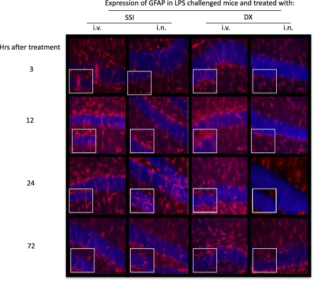
Figure 3.

Effect of dexamethasone (DX) on glial fibrillary acidic protein (GFAP) expression in lipopolysaccharide (LPS)‐treated mice. Representative immunofluorescence of 30 μm coronal sections of mouse brain of the different groups stained with anti‐GFAP antibodies (red) and 4',6‐diamidino‐2‐phenylindole (DAPI) (blue nuclei). The pictures derive from LPS‐injected mice, treated intranasally (i.n.) or intravenously (i.v.) with saline or DX 3, 12, 24 and 72 h later. The highest number of astrocytes with morphological characteristics of activated cells was observed 24 h after DX treatment. Bottom images represent a fivefold magnification of the region outlined in the box in the corresponding upper image. [Colour figure can be viewed at wileyonlinelibrary.com]
