
Figure 6.
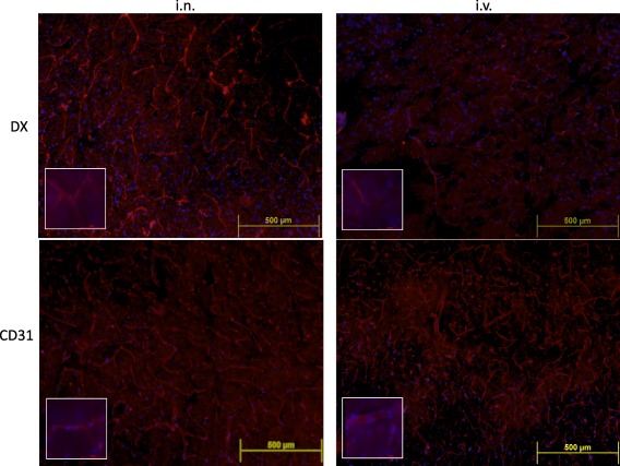
Representative immunofluorescence images of brain slices (four to five mice per group) obtained 24 h after intranasally (i.n.) or intravenously (i.v.) dexamethasone (DX) administration. Merged images of DX (red) or CD31 (red), and 4',6‐diamidino‐2‐phenylindole (DAPI) (blue) staining are shown at ×10 (upper panel) and ×100 (bottom panel) magnification, respectively. Brain vessels in the cortex were distinguished more clearly in i.n. DX‐treated (4·82 pixels/µm2) than in brains of i.v. DX‐treated (1·97% pixels/µm2) mice. [Colour figure can be viewed at wileyonlinelibrary.com]