Figure 5.

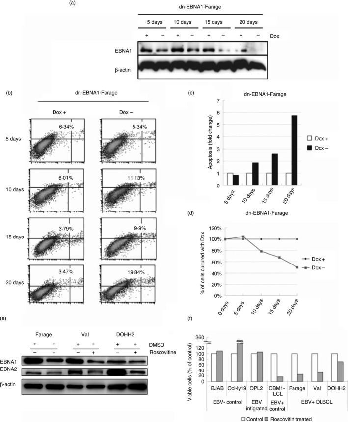
Epstein–Barr virus (EBV) provides survival factors to EBV‐positive diffuse large B‐cell lymphoma (DLBCL) lines. (a) Immunoblot analysis of total cell extracts of dominant negative EBV nuclear antigen 1 (dnEBNA1) ‐Farage cultured with or without 1 μg/ml doxycycline (Dox) for 5, 10, 15 and 20 days with β‐actin and EBNA1 antibodies. (b) dn‐EBNA1‐Farage cells were cultured with or without 1 μg/ml Dox for 5, 10, 15 and 20 days and apoptosis was assayed using Annexin V‐FITC and propidium iodide (PI) according to the manufacturer's instructions. The x‐axis of dual parametric dot plots represent PI fluorescence and y‐axis represent Annexin V‐FITC fluorescence. Cells were considered ‘apoptotic’ if positive for Annexin V. (c) Bar charts showing the relative percentage of apoptotic dnEBNA1‐Farage cells (Annexin V+) cultured with Dox for 5, 10, 15 and 20 days in comparison with the cells cultured without Dox. The result is presented as relative fold change of the percentage of apoptotic cells where the control cultured with Dox was arbitrarily defined as 1. (d) Relative viable dn‐EBNA1‐Farage cells (Annexin V‐) cultured without Dox for 5, 10, 15 and 20 days in comparison with the control cells cultured without Dox. The result is presented as a percentage of the control cells where control cells cultured with Dox, arbitrarily defined as 100%. (e) Immunoblot analysis of total cell extracts of Farage, Val and DOHH2 treated with 3 μg/ml Roscovitine for 12 days with β‐actin EBNA2 and EBNA1 antibodies. (f) Relative viable cells of the Roscovitine (3 μg/ml, 12 days)‐treated Farage (EBV + DLBCL), Val (EBV + DLBCL), DOHH2 (EBV + DLBCL), OCI‐Ly19 (EBV – DLBCL), BJAB (EBV – lymphoma), OPL2 (EBV‐integrated DLBCL) and CBM1 lymphoblastoid cell line (EBV + LCL) as measured by dye‐exclusion assay. The result is presented as percentage of the control cells treated with DMSO as the values of control cells were arbitrarily defined as 100%.
