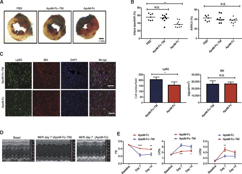
Fig. 6. ApoM-Fc administration attenuates I/R injury in the heart.
WT mice were administered PBS (n = 7 mice) or either ApoM-Fc (4 mg/kg) (n = 9 mice) or ApoM-Fc–TM (4 mg/kg) (n = 9 mice) after I/R injury. (A) Representative images of left ventricular (LV) slices with Alcian blue and 2,3,5-triphenyltetrazolium chloride (TTC) staining (red), which indicates viable myocardium. (B) Quantitative measurement of area at risk (AAR)/LV area and infarct/AAR area was performed in a blinded manner. Median values (one-way nonparametric ANOVA followed by Tukey’s test); **P < 0.01; N.S. was P > 0.7 (left graph) and P > 0.27 (ApoM-Fc) and P > 0.97 (ApoM-Fc–TM) (right graph). (C) Heart sections were stained for Ly6G and IB4, and neutrophils and capillary density were quantified for pixel density. n = 9 mice per treatment; mean ± SEM. P < 0.03 (left graph) and P > 0.85 (right graph), unpaired two-tailed parametric t test. DAPI, 4′,6-diamidino-2-phenylindole. (D) Representative images of two-dimensional (2D) guided M-mode echocardiography of the LV at baseline and 7 days after MI/R injury. n = 6 mice per treatment. (E) LV end-diastolic (LVDd) diameter, LV end-systolic (LVDs) diameter, and fractional shortening (FS) of (D) were measured at the indicated time points after MI/R injury. n = 6 mice per treatment; means ± SEM. Two-way ANOVA and Sidak’s multiple-comparisons test; *P < 0.05; **P < 0.01; ***P < 0.005; ****P < 0.001.

