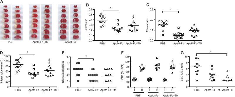
Fig. 7. ApoM-Fc administration attenuates I/R injury in the brain.
(A) WT mice were subjected to 60 min of focal cerebral ischemia by MCAO. Right after reperfusion, mice received ApoM-Fc (4 mg/kg) (n = 11 mice), ApoM-Fc–TM (4 mg/kg) (n = 11 mice), or PBS (n = 10 mice) by intraperitoneal injection. Representative images of TTC staining of seven, 1-mm-thick brain coronal slices 23 hours after reperfusion. Two mice per group are shown. (B and C) Infarct (B) and edema (C) ratios were calculated by image analysis and reported as a ratio of the non-ischemic hemisphere. Infarct ratios were corrected for edema. (D) Total infarct volume in cubic millimeters, corrected for edema. (E) Neurological deficit scores were assessed 23 hours after reperfusion. (F) Relative cerebral blood flow (rCBF) in the MCA territory was measured by laser speckle flowmetry during MCAO surgery. rCBF during occlusion (I) and after reperfusion (R) is shown. CL, contralateral. The individual values and the median are shown. *P < 0.05 (one-way nonparametric ANOVA followed by Dunn’s test). (G) Evans blue dye (EBD) extravasation into the brain after MCAO stroke was quantified as described. IL, ipsilateral. Individual values and mean ± SEM are shown from n = 8 to 10 mice per treatment. P < 0.05, one-way ANOVA nonparametric test.

