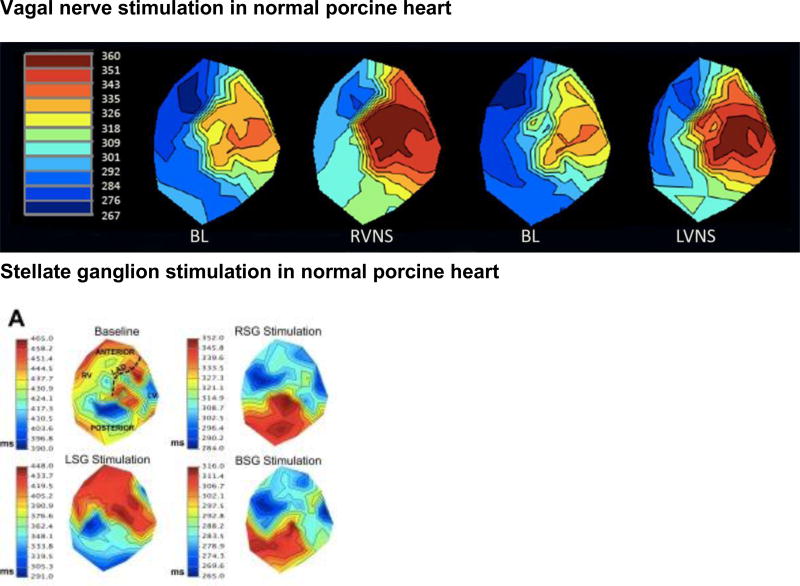
Figure 7.
A, Activation recovery interval (ARI) maps at baseline and during right (RVNS) and left (LVNS) vagal nerve stimulation in a normal porcine heart. No significant regional differences in responses were found. Adapted from Yamakawa et al.79 B, ARI maps in control porcine hearts at baseline (BL) and during right (RSG), left (LSG) and bilateral (BSG) stellate ganglion stimulation. Myocardial regions are displayed in the BL control map. Adapted from Ajijola et al.130

