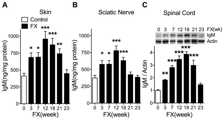
Fig. 7. Changes in IgM levels in the ipsilateral hindpaw skin, sciatic nerve, and lumbar spinal cord at 3, 7, 12, 18, 21, and 23 weeks after fracture (FX) in wildtype mice.
IgM levels were determined by enzyme immunoassay in skin (A) and sciatic nerve (B) and by western blot analysis in spinal cord (C), due to the low IgM levels observed in the spinal cord. After fracture there was a gradual increase in IgM levels in the hindpaw skin (A), sciatic nerve (B), and spinal cord (C), peaking at between 12 and 18 weeks post-fracture and then declining to control levels by 23 weeks post-fracture. Data were analyzed using a one-way analysis of variance followed by a Holm-Sidak test for post hoc contrasts, error bars indicate SEM. *P< 0.05, **P< 0.01, and ***P< 0.001 for fracture (FX, n=4–8) vs control (n=4–8). Control: nonfracture wildtype mice, FX: fracture wildtype mice, FX(week): weeks after fracture.

