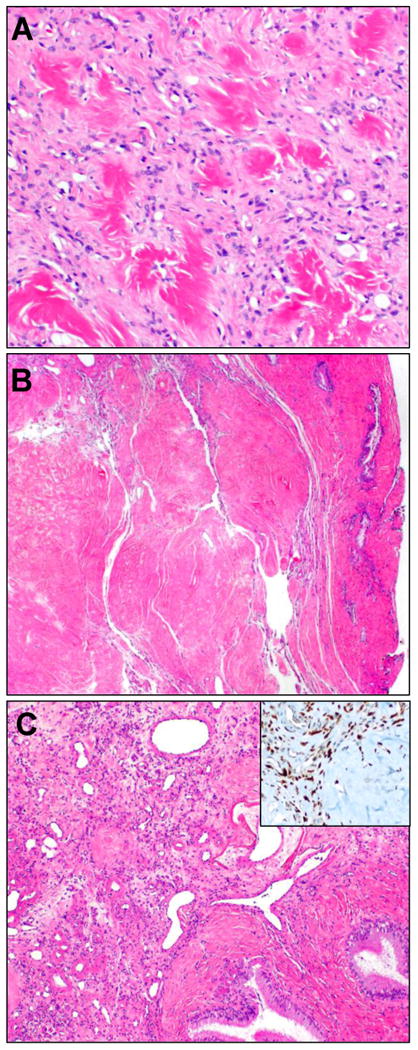
Figure 6.

Prominent stromal hyalinization, in association with vessels and in the stroma, imparted the appearance of collagen rosettes (A) with the appearance of amianthoid fibers or keloidal collagen. In one case, this feature predominated (B), such that a nodular amyloidoma had entered the differential. Upon close inspection, distinctive SFT vasculature was apparent (C), and STAT6 was positive (inset).
