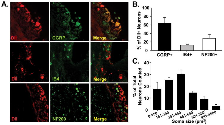
Figure 4. Predominantly peptidergic primary afferent neurons innervate the tongue.
Tongue afferent neuron labeling using the lipophilic retrograde tracer, DiI. A) Representative images from co-staining of DiI-positive (red) TG sections with CGRP (top, green), IB4-FITC (middle, green), and NF200 (bottom, green). Co-expression is indicated by yellow overlap (Merge). B) Pooled data from 3 female TG pairs indicated that DiI-positive neurons had predominantly more CGRP-immunolike reactivity (IR) (64.3 ± 13.2%) compared to IB4-IR (13.2 ± 1.6%) and NF200-IR (29 ± 8.9%) populations. C) Histogram of neuronal size distribution of cells retrogradely labeled from the tongue. Tongue afferent neurons are predominately small to medium (soma size 151–450 μm2).

