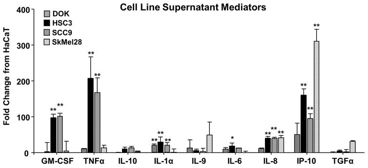
Figure 6. Analysis of cell line supernatant protein contents revealed multiple pro-inflammatory and pro-nociceptive mediators.
Cytokines present in supernatants of cancer cell lines (HSC3, SCC9) as well as precancerous dysplastic oral keratinocytes (DOK) cell line, melanoma (SkMel28) cell line, and immortalized non-tumorigenic keratinocytes (HaCaT) cell line were quantitated using MILLIPLEX® MAPmultiplex cytokine biomarker magnetic bead panel for detection of 38 mouse cytokines and chemokines. Pooled data demonstrate that of the 38 mediators measured, 9 had a ≥ 10-fold change in DOK (gray), HSC3 (black), SCC9 (black stripe), or SkMel28 (gray stripe) compared to HaCaT. Two-way ANOVA demonstrated a significant interaction between cell lines tested and mediators measured (p < 0.01). Dunnett’s post hoc analysis is represented on the graph (* p < 0.05, ** p < 0.01).

