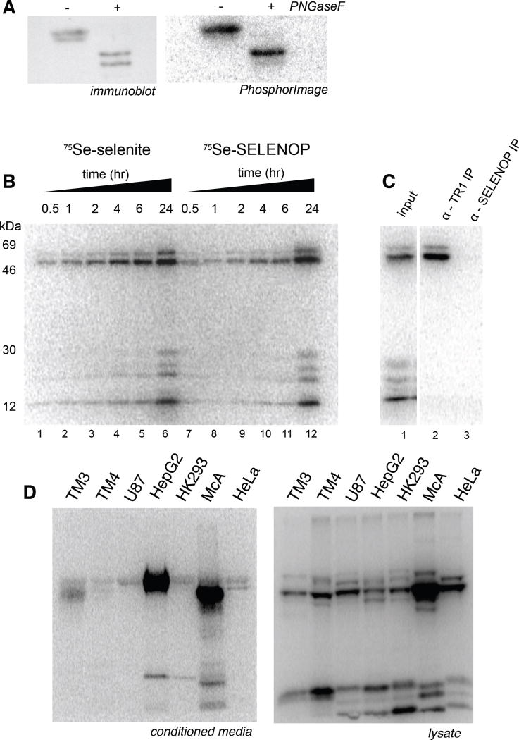
Figure 1. 75Se-SELENOP can be used as a selenium source for intracellular selenoprotein production.
(A) Conditioned medium from HepG2 cells grown in the presence of 75Se-selenite was concentrated ~50-fold and a sample was treated with PNGaseF. The samples were resolved by SDS-PAGE and SELENOP was detected by immunoblot (left) and phosphorimage analysis (right). (B) HeLa cells were incubated with ~2 pmol of 75Se-SELENOP or 100 nM 75Se selenite for the time points indicated then switched to normal growth medium and allowed to grow for a total of 24 hours. Endogenous selenoproteins that utilized the 75Se were resolved by SDS-PAGE and detected by PhosphorImage analysis. (C) HeLa cells were labeled with 75Se-SELENOP as described in (A) and lysates were incubated with either an anti-thioredoxin reductase or anti-SELENOP antibody. Immunoprecipitated 75Se-labeled proteins were resolved by SDS-PAGE and detected by PhosphorImage analysis. (D) 75Se-selenite was used to label the endogenous selenoproteins in the cell types indicated: mouse Leydig cells [TM3], mouse Sertoli cells [TM4], human glioblastoma [U87], human hepatoma [HepG2], rat hepatoma [McArdle 7777], human epithelial [HeLa]. After 24 hours of labeling, 4% of the media (left) and 10% of the lysate (right) was resolved by SDS-PAGE and detected by PhosphorImage analysis.

