Figure 1.
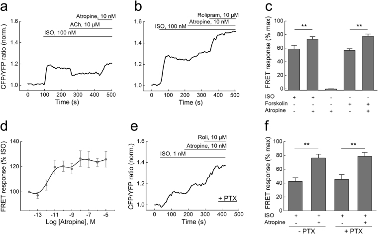
Single-cell FRET analysis of intracellular cAMP levels in adult ventricular mouse cardiomyocytes transgenically expressing the Epac1-camps sensor. (a) The β-AR agonist isoproterenol (ISO, 100 nM) increases intracellular cAMP (indicated by normalized CFP/YFP ratio), and this response is partially reversed by acetylcholine (ACh, 10 µM). The ACh effect is completely blocked by atropine (10 nM), and the cAMP levels are even further increased compared to the steady-state reached after ISO stimulation. CFP, enhanced cyan fluorescent protein; YFP, enhanced yellow fluorescent protein. (b) Atropine increases cAMP levels beyond the plateau reached after ISO stimulation. They can be further elevated by the selective PDE4 inhibitor rolipram (10 µM). Data in a and b are representative traces, quantification is shown (c) as mean ± s.e.m. (n = 5–8). The data are presented as a % of the maximal FRET response reached after ISO/rolipram or forskolin/rolipram stimulation. (d) Concentration-response dependency of atropine on cAMP levels in cardiomyocytes after ISO prestimulation. (e) Inhibition of Gi-proteins with pertussis-toxin (1.5 µg/ml for 7–8 h) does not affect the atropine mediated stimulation of cAMP. Here we used 1 nM ISO, since 100 nM lead to a complete saturation in PTX-treated cells. Representative experiment (n = 5). Quantification of the FRET ratio changes is shown in (f). Here and in C: **differences are statistically significant at, p < 0.01 by one-way ANOVA.

