Figure 3.
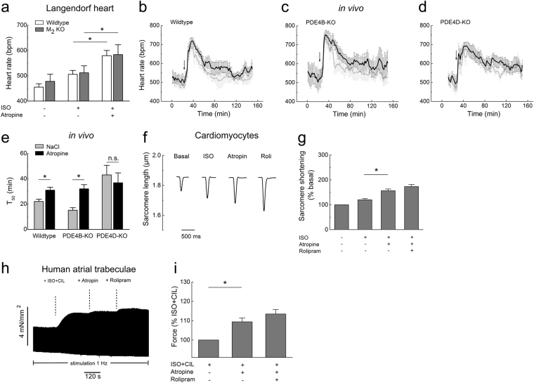
Atropine augments cardiac function in PDE4 dependent and muscarinic receptor independent manner. (a) Heart rate measurements in wildtype and M2-receptor knockout Langendorff hearts perfused with ISO alone (10 nM) or with ISO plus atropine (10 nM). Atropine applied after ISO significantly increases the beating frequency in both genotypes (n = 5). *p < 0.05, by paired t-test. (b–e) Averaged heart rate tracings and T50 values obtained from in vivo telemetry experiments in wildtype vs PDE4B or PDE4D knockout mice injected with atropine (0.5 mg/kg, denoted by arrow, black traces) or saline (NaCl, grey traces) control indicate that PDE4D but not PDE4B is involved in the hydrolysis of cAMP which regulates the duration of atropine-induced heart rate increase. T50 was defined as the duration of the increase in heart rate measured from half-maximal increase to half-maximal return to baseline. Number of mice used was 8, 5 and 4 for wildtype, PDE4B-KO and PDE4D-KO, respectively. (f) Representative traces from single cardiomyocyte contractility measurements by edge-detection show a positive inotropic effect of atropine (10 nM) applied after ISO (3 nM). Quantification is in (g), n = 10–13. In (e) and (g), *denotes significant differences p < 0.05 by one-way ANOVA. n.s. – not significant. (h) Original representative trace showing the effect of atropine on the developed force of contraction in human right atrial trabeculae. Atropine increases the force of contraction in trabeculae which were prestimulated with 1 nM ISO and 10 µM cilostamide (CIL). (i) Quantification of the contractility data. The data for each experiment were normalized to the force developed after prestimulation with ISO plus CIL. Mean ± s.e.m. (n = 5).*The differences are statistically significant (p < 0.05, by paired t-test).

