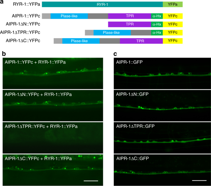
Fig. 7.
Bimolecular fluorescence complementation between AIPR-1 and RYR-1 in vivo. a Schematic diagrams showing the various fusion proteins used in the bimolecular fluorescence complementation assays. b Deletion of the TPR domain but not any other parts of AIPR-1 prevented AIPR-1::YFPc from reconstituting YFP fluorophore with RYR-1::YFPa in ventral cord motor neurons. c GFP-tagged AIPR-1 with the various deletions showed comparable expression levels in ventral cord motor neurons. Scale bars, 20 µm

