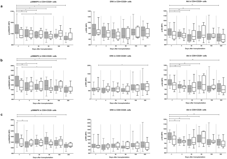
Figure 3.
P-p38MAPK, p-ERK and p-Akt in T cell subsets of TAC- (grey) and BELA- (white) treated patients after stimulation with PMA/ionomycin. (a) p-p38MAPK (left), p-ERK (middle) and p-Akt (right) within CD4+CD28+ T cells. P-p38MAPK and p-Akt, but not p-ERK, were significantly decreased in TAC-treated patients at different time points after transplantation, in contrast to BELA-treated patients. (b) p-p38MAPK (left), p-ERK (middle) and p-Akt (right) within CD8+CD28+ T cells. P-p38MAPK and p-Akt, but not p-ERK, were inhibited when TAC was given. BELA caused an increase in p-ERK at day 360. (c) p-p38MAPK (left), p-ERK (middle) and p-Akt (right) within CD8+CD28− T cells. Phosphorylation inhibition by TAC is comparable with CD8+CD28+ T cells. (Data are plotted as box and whiskers indicating total range; n = 20 TAC-treated patients and n = 20 BELA-treated patients) *p < 0.05, **p < 0.01, ***p < 0.001.

