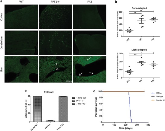
Fig. 3.

Histological and clinical parameters of F42. (a) Representative images show AFSM accumulation in PPT1−/− brains (cortex and cerebellum). In contrast, AFSM was not detected in the cortex or cerebellum of WT or F42 animals. Representative images show similar levels and distribution of AFSM in the livers of PPT1−/− and F42 animals. No AFSM was detected in WT livers. All images were taken at the same magnification of 20x (100 μm). (b) Electroretinography of F42 and control eyes at 7 months. There was a significant decrease in b-wave amplitudes in the light-adapted (WT p < 0.0001, F42 p < 0.01) and dark-adapted (WT p < 0.001, F42 p < 0.001) PPT1−/− mice compared to WT and F42 mice. There was no significant difference between F42 and WT b-wave amplitudes in light-adapted or dark-adapted conditions. (c) Rotarod analysis of F42 mice showed no deficit in motor function at 7 months of age. PPT1−/− animals could not stay on the rotarod at 7 months of age. However, 7-month-old F42 mice were able to stay on the rotarod for 60 s. Similarly, the WT mice were also able to stay on for the duration of the study. (d) The lifespan of F42 mice (orange) was not reduced compared to WT animals (black) out to 1 year. In contrast, PPT1−/− animals had a median lifespan of 238 days (blue)
