Fig. 3.

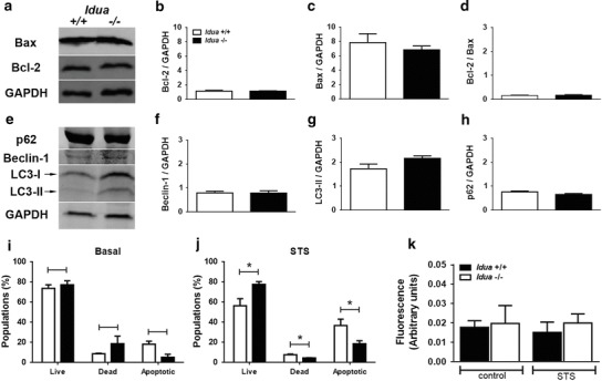
Idua −/− fibroblasts are more resistant to apoptotic induction. Total protein extracts of Idua +/+ and Idua −/− fibroblasts (30–50 mg) were loaded into 10–15% SDS-polyacrylamide gels for evaluation of basal apoptotic (Bcl-2 and Bax) and autophagic (p62, beclin-1 and LC3-II) markers. No differences were observed in Bcl-2 and Bax relative expression (a–d), as well as in autophagy markers such p62, beclin-1, and LC3-II (e–h). Moreover, no changes in basal viability of peritoneal Idua −/− fibroblasts were observed (i). However, incubation with STS (5 μM) for 4 h resulted in an increase of viable (live) and a decrease of dead and apoptotic cells (j). In addition, no differences were observed in caspase activity of control (no STS) and STS-treated cells (k). Data are expressed as the mean ± S.E.M. of three independent experiments. P < 0.05 (Student’s t test). n = 4–6
