Figure 4.
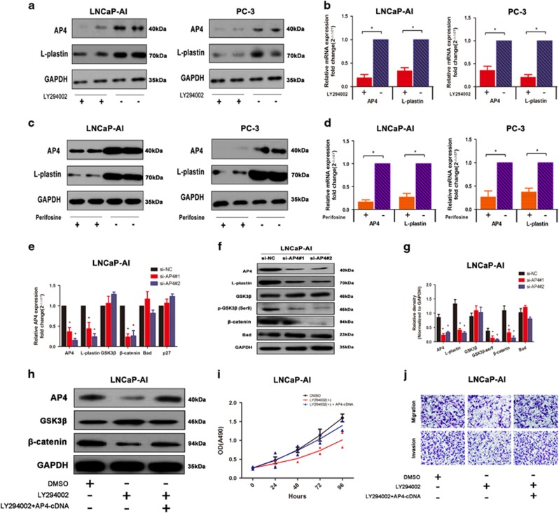
AP4 regulates PCa via activation of PI3K/AKT pathway. (a,b) LNCaP-AI and PC-3 cells were treated with PI3K inhibitor LY294002, AP4 and L-plastin mRNA and protein levels were determined by qRT-PCR and western blot analysis. GAPDH was used as loading control. (c,d) LNCaP-AI and PC-3 cells were treated with AKT inhibitor Perifosine, AP4 and L-plastin mRNA and protein levels were determined by qRT-PCR and western blot analysis. (e–f) The expressions of AP4, L-plastin, GSK3β, β-catenin and Bad after transfection with si-NC, si-AP4#1 and si-AP4#2 were examined by qRT-PCR and western blot analyses in LNCaP-AI cells. (g) Relative density of AP4, L-plastin, GSK3β, β-catenin and Bad protein expressions after normalization to GAPDH in LNCaP-AI cells. (h–j) The levels of AP4, β-catenin and GSK3β were examined with the PI3K inhibitor LY294002, and overexpression of AP4 could partly rescue the inhibitory effects in the changes of AP4 and β-catenin in western blotting analysis, MTT assay and transwell assays. Error bars indicate S.D.s (n=3), *P<0.05. **P<0.01

