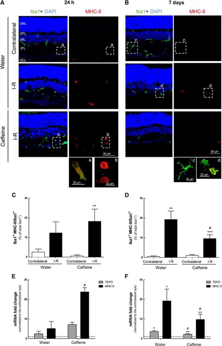
Figure 5.
Effect of caffeine administration in microglia reactivity elicited by I–R injury. Caffeine (1 g/l) was administered in the drinking water for 2 weeks prior ischemic injury and until the end of the experiment (24 h and 7 days of reperfusion). (A and B) Microglia reactivity was assessed in retinal sections by labeling with major MHC-II (reactive microglia, red) and Iba1 (general marker, green) at 24 h (a) and 7 days (b) of reperfusion. Nuclei were stained with DAPI (blue). Representative images are depicted. Note in the inserts (a–d) the alteration of microglia morphology from ramified to ameboid morphology, indicating microglia reactivity. (C and D) The number of reactive microglia/macrophages (MHC-II+Iba1+ cells) was expressed as the percentage of total number of microglia/macrophages (Iba1+ cells) in the retinal section at 24 h (c) and 7 days (d) of reperfusion. **P<0.01, ***P<0.001, significantly different from contralateral eye; #P<0.05, significantly different from I–R retinas of water-drinking animals, Mann–Whitney test. (E and F) The mRNA expression of the markers of microglia activation, TSPO and MHC-II, was assessed by qPCR after 24 h (e) and 7 days (f) of reperfusion. Results are presented as fold change of the contralateral eye. *P<0.05, **P<0.01, significantly different from contralateral eye, Wilcoxon's signed-rank test; #P<0.05, significantly different from water-drinking group, Mann–Whitney test. ONL, outer nuclear layer; OPL, outer plexiform layer; INL, inner nuclear layer; IPL, inner plexiform layer; GCL, ganglion cell layer

