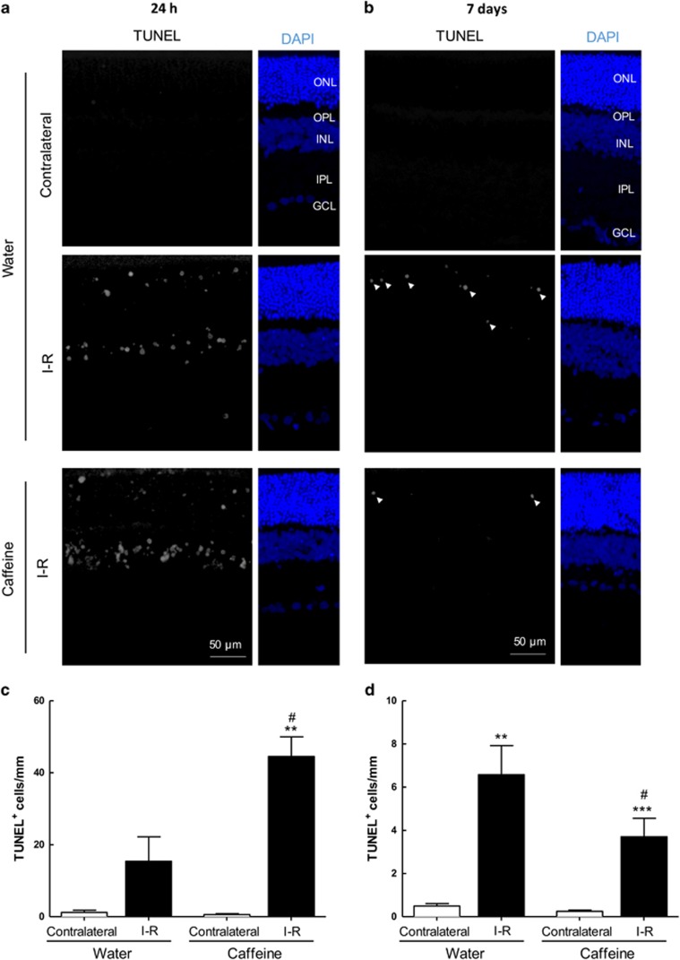
Figure 6.
Effects of caffeine administration in cell death induced by transient retinal ischemia. Caffeine (1 g/l) was administered in the drinking water for 2 weeks prior injury and until the end of the experiment (24 h and 7 days of reperfusion). (a and b) Cell death was assayed in retinal cryosections by TUNEL assay at 24 h (a) and 7 days (b) of reperfusion. Nuclei were stained with DAPI (blue). Representative images are depicted. (c and d) TUNEL+ cells (gray, some TUNEL+ cells are indicated with arrowheads) were counted and were expressed per mm of retina. **P<0.01 and ***P<0.001, significantly different from contralateral eye; #P<0.05, significantly different from I–R retinas of water-drinking animals, Mann–Whitney test. ONL, outer nuclear layer; OPL, outer plexiform layer; INL, inner nuclear layer; IPL, inner plexiform layer; GCL, ganglion cell layer

