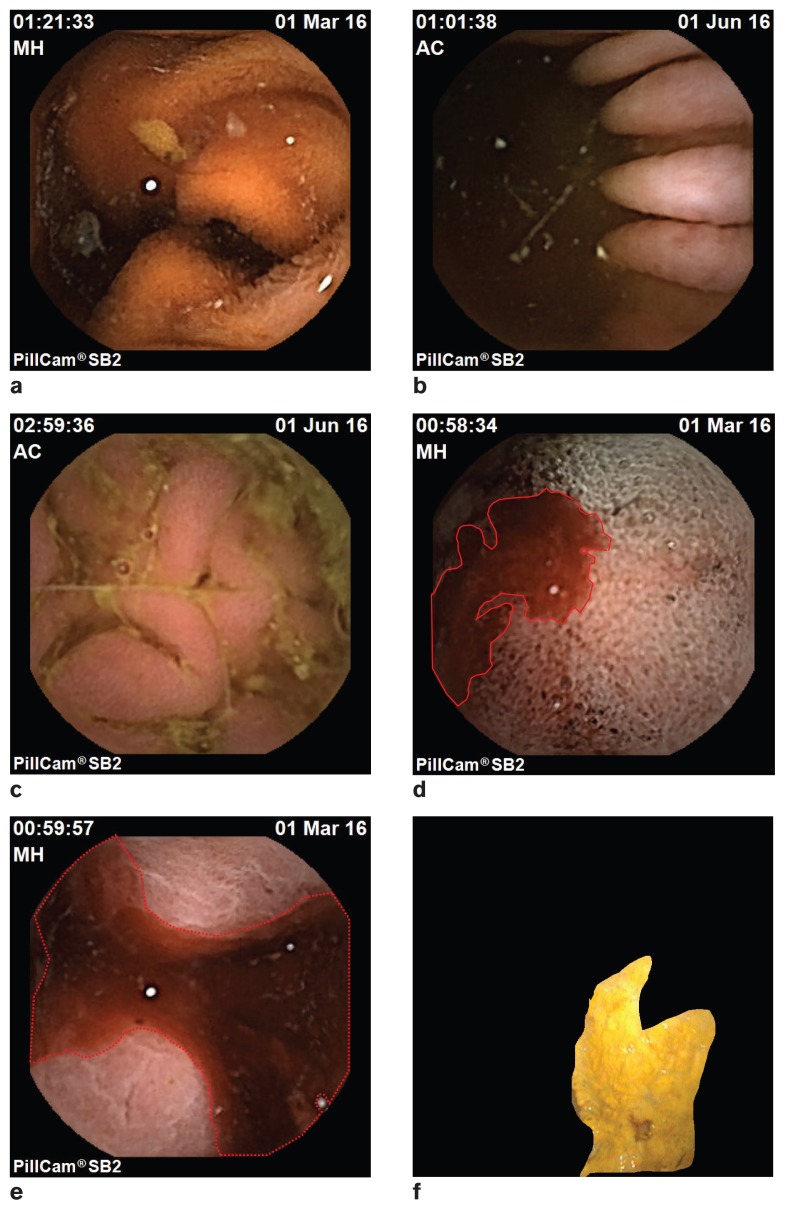
Figure 2.
a to c — Images from the PillCam at various regions of the stomach and small intestine. d to e — PillCam image indicating presence of blood (marked using an auto-detection algorithm during offline diagnosis); f — Regular gastroscopy image with an ulcerous region marked (using automated segmentation algorithm during offline diagnosis).

