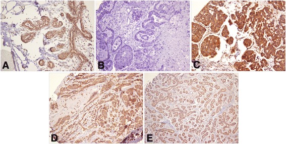
Fig. 1.

Granular cytoplasmic expression of leptin in breast cancer. a strong positive staining in normal breast tissue (20 X); b negative stained breast cancer (20 X); c strong positive staining in epithelial cells of breast cancer (20 X); d weak positive staining in epithelial cells of breast cancer (20 X); e weak positive staining in fibroadenoma (10 X)
