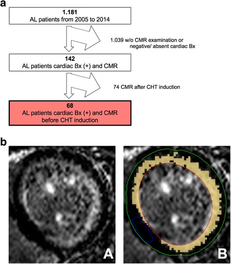
Fig. 1.

a Patient selection flow chart. From 2005 to 2014 a total of 1.181 AL patients visited our institution. Patients had to be excluded for negative or absent cardiac biopsy (Bx), absent CMR examination, or CMR performed after induction of chemotherapy (CHT). The final study cohort consists of 68 AL patients. b Late gadolinium enhancement (LGE) quantification by semiautomatic 5 SD-threshold selection. Contours for endo- (red) and epicardial borders (green) as well as for unenhanced reference myocardium (blue) were drawn manually. Signal intensities above 5-fold standard deviation of the reference myocardium were accounted to the LGE volume in relation to unenhanced myocardium (relative LGE, orange)
