
Fig. 3.
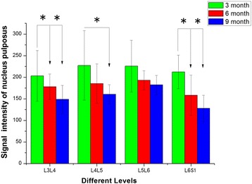
The signal intensity of the nucleus pulposus in different levels of the T2-weighted magnetic resonance images. *Indicates significant differences between different months, P < 0.05. No significant differences were observed in the intervertebral discs of different levels between 6- and 9-month-old groups (L3–L4 represent the intervertebral disc between the third and the fourth lumbar vertebra, and so on)