
Fig. 2.
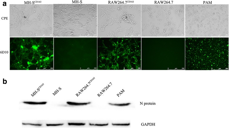
PRRSV infections in MH-SCD163 and RAW264.7CD163 cell lines. The MH-S and RAW264.7 cell lines and PAMs were inoculated with JXA1 at 1 MOI. a CPEs were visualized at 24 hpi using an inverted microscope (200×). Meanwhile, cells were fixed and permeabilized to measure virus infection using immunofluorescence staining of virus using anti-PRRSV N protein-specific mAb (6D10). Images are representative one of three independent experiments. b Cell infection was detected using anti-PRRSV N protein-specific mAb labeling of western blot using GAPDH as the control