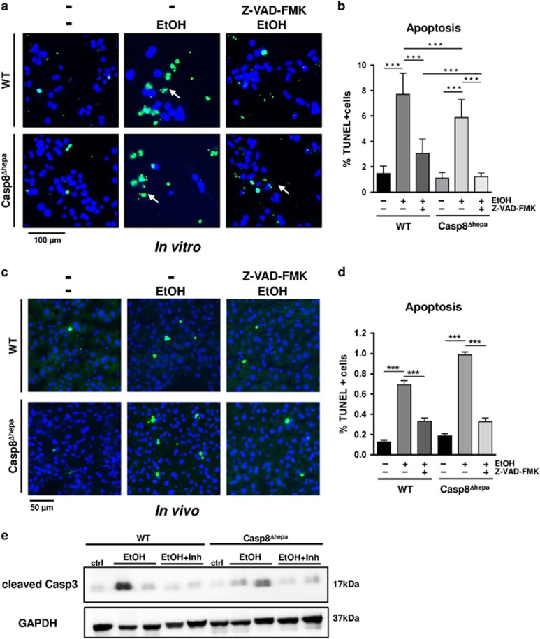
Figure 6.
Pan-caspase inhibitors prevent alcohol-induced hepatic apoptosis in vitro and in vivo. (a–b) In vitro analysis. Primary hepatocytes from WT and Casp8Δhepa mice were isolated, plated and stimulated with 100 mM EtOH for 24 h with or without pan-caspase inhibitor Z-VAD-FMK (a) TUNEL staining. Apoptotic nuclei are stained in green. Total nuclei are counter-stained with DAPI (blue). (b) Quantification of cellular apoptosis. Data were calculated as percentage of TUNEL-positive cells per magnification field. (c–e) In vivo analysis. WT and Casp8Δhepa mice (n=4–6) were fasted for 6 h and then injected (i.v.) with 20 μg/g body weight of Z-VAD-FMK/8% or solvent control. Subsequently, they were fed with 30% (w/v) EtOH by three equally divided oral gavages in 20-min intervals. All animals were killed 12 h after last EtOH feeding. (c) TUNEL staining of liver cryosections. (d) Quantification of apoptosis. For each animal, 10 independent magnification fields (× 200) were counted. (e) Immunoblot analysis of Caspase-3 activation

