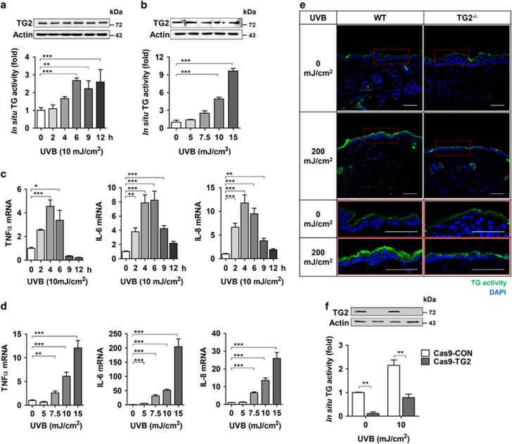
Figure 3.
UV irradiation activates keratinocyte TG2. (a and b) Levels of TG2 protein and in situ TG activity in HaCaT cells were determined by western blot analysis and biotinylated pentylamine (BP) incorporation assay, respectively. HaCaT cells were exposed to UV irradiation (a, 10 mJ/cm2, n=4; b, doses indicated, n=4) and collected at the time indicated (a) or after 6 h of UV irradiation (b). (c and d) mRNA levels of TNF-α, IL-6, and IL-8 were determined by QRT-PCR in HaCaT cells exposed to UV irradiation as (a) and (b) (n=3). (e) In situ TG activity in the skin from UV-irradiated WT and TG2−/− littermates (n=3). (f) In situ TG activity in TG2-deficient HaCaT cells (Cas9–TG2) after 6 h of UV irradiation (10 mJ/cm2). All data are represented as mean±SEM. *P<0.05; **P<0.01; ***P<0.001

