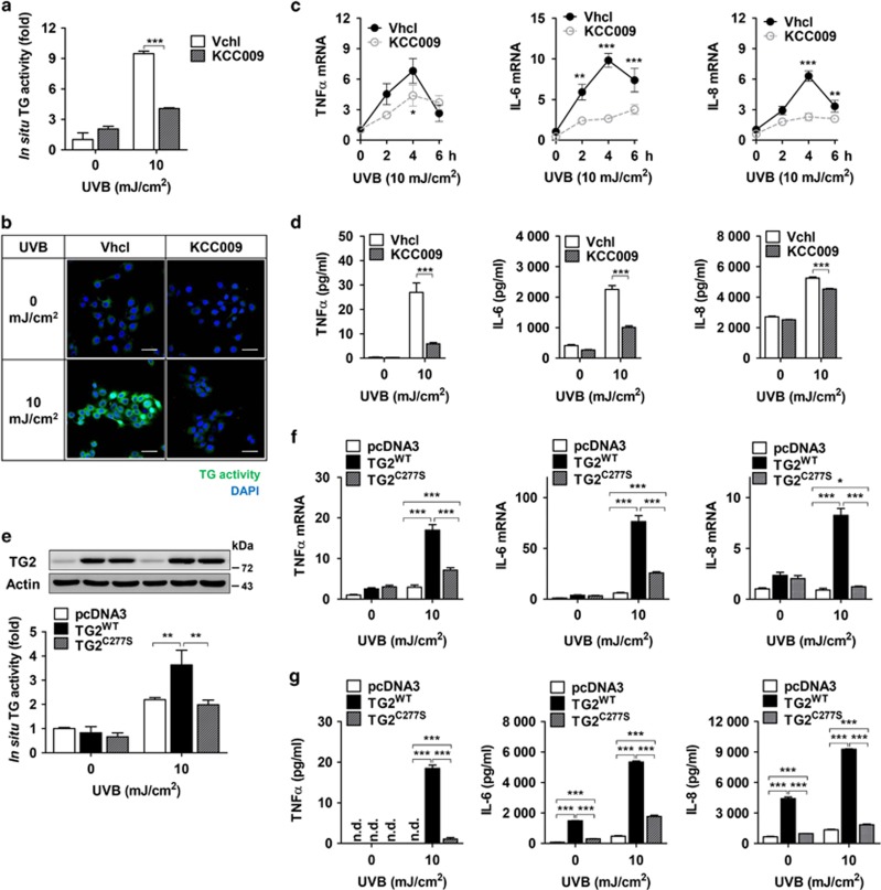
Figure 4.
TG inhibition suppresses the production of cytokines in UV-irradiated keratinocytes. (a and b) HaCaT cells were pretreated with KCC009 (250 μM) or vehicle (Vhcl) 1 h before UV irradiation (10 mJ/cm2). The cells were incubated for 6 h after UV irradiation in the same media. In situ TG activity was measured by the BP incorporation assay (a, n=3) and visualized by confocal microscopy (b). Scale bars, 50 μm. (c and d) Levels of mRNA (c, n=3) and proteins (d, n=3) for TNF-α, IL-6, and IL-8 were measured in KCC009-pretreated and UV-irradiated HaCaT cells at the times indicated (c) or 6 h after UV irradiation (d). (e) Levels of TG2 protein and in situ TG activity were measured in HaCaT cell lines expressing pcDNA3, WT (TG2WT), or active-site mutant TG2 (TG2C277S) established by transfection and selection (n=4). (f and g) Levels of mRNA (f, n=4) and proteins (g, n=4) for TNF-α, IL-6, and IL-8 were measured in HaCaT cell lines expressing pcDNA3, WT (TG2WT) or active-site mutant TG2 (TG2C277S) at 6 h after UV irradiation. n.d., not determined. All data are represented as mean±SEM. *P<0.05; **P<0.01; ***P<0.001

