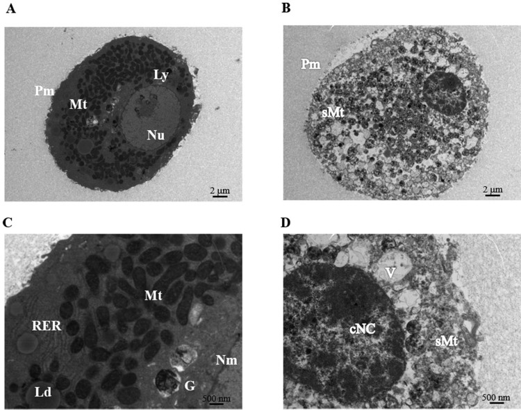
Fig. 5.
Transmission electron microscopy (TEM) of hepatocytes within microbeads after 24-h culture. Representative TEM images of normal healthy hepatocytes (left panel) and necrotic/apoptotic hepatocytes (right panel). (A, B) at magnification of 1,400×, and (C, D) at high magnification of 4,800×. cNC, condensed nuclear chromatin; G, Golgi apparatus; Ld, lipid droplet; Ly, lysosomes, Mt, mitochondria; N, nuclei; Nu, nucleoli; Nm, nuclear membrane; Pm, plasma membrane; RER, rough-surfaced endoplasmic reticulum; sMt, swelling mitochondria; V, vacuole.

