Figure 1.
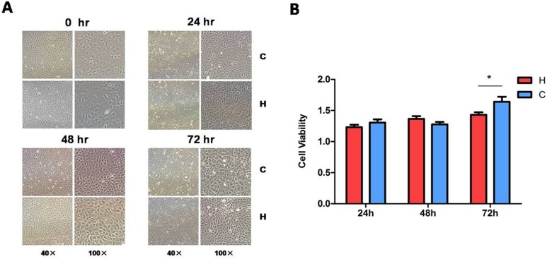
Morphology and viability of the rat kidney epithelial cells (NRK-52E) exposed to hypoxia. After the cells were exposed to hypoxia at 1% O2 for 24, 48, or 72 h, cell morphology was examined by light microscopy (A) and the cell viability was measured by 3-[4,5-dimethylthiazol-2-yl]-5-[3-carboxymethoxypheny]-2-[4-sulfophenyl]-2H-tetrazolium (MTS) assay. (B) At least 3 independent experiments were carried out in all groups. H, hypoxia; C, normoxic control. The photomicrographs were taken with 40× magnification (scale bar, 100 μm) and 100× magnification (scale bar, 40 μm). *P < 0.05. Note that cell morphology under light microscopy showed no appreciable difference between the control and hypoxia, while the MTS assay indicated a slight decrease in cell viability after 72 h of hypoxia.

