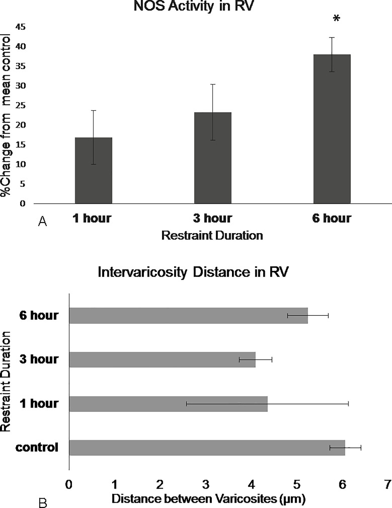
Fig 7. NOS in the DRN rostral ventromedial region show differences in activation following restraint stress.
(A) NOS activity barely shifts after 1 hour of restraint then increases following 3 hours of restraint and significantly increases following 6 hours of restraint. (B) Intervaricosity spacing decreases following 3 hours of restraint but by 6 hours, the intervaricosity spacing is closer to control values. Percentages were calculated by subtracting each animal per experimental group by the mean of the control then dividing by the control mean and multiplying by 100. Significance is denoted by an asterisk and compared to the control. Error bars are representative of standard error.

