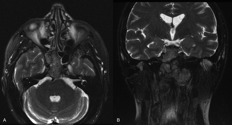
Fig. 1.

Preoperative gadolinium-enhanced T2-weighted ( A ) axial and ( B ) coronal magnetic resonance imaging sections. An enhanced clival mass, 48 × 36 × 55 mm, is shown, invading the right sphenoid sinus, the occipital condyles, the dens, and the anterior arch of C1. Radiological features were consistent with chordoma.
