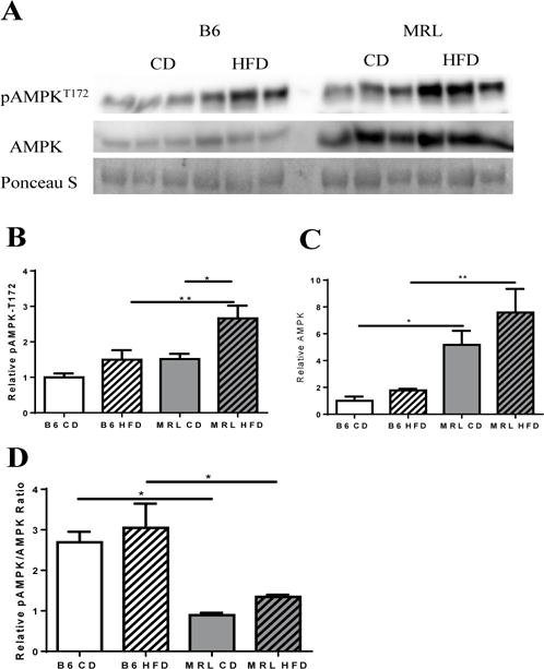
Figure 1.

Hepatic expression of AMPK and pAMPKT172. (A) Western blot image of AMPK and pAMPKT172 in liver tissue lysates of each mouse group (normalized to total protein content derived from Ponceau S stain). (B) Densitometry analysis reveals significantly increased pAMPKT172 expression in MRL HFD with no significant changes on their B6 age-pair controls. (C) Densitometry analysis of AMPK reveals significantly increased AMPK in MRL vs B6, with the HFD-fed MRL group expressing a ~5-fold difference when compared with diet-matched B6. (D) Overall changes in the pAMPKT172/AMPK ratio, with both strains showing a marked difference against their diet-matched cohorts. n=5–6; * p <0.05, ** p<0.01. AMP dependent protein kinase (AMPK), phosphorylated AMPK (pAMPKT172).
