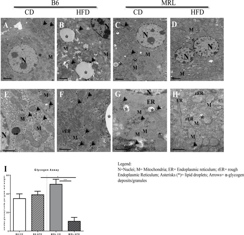
Figure 3.

MRL mice are resistant to hepatic fat accumulation and exhibit diminished α-glycogen deposits when subjected to a HFD. (A–H) Transmission electron microscopy images of mice liver biopsies. (A-D) Upper panel shows representative images of lipid deposition in each group at ×10,000 magnification. In contrast to (A) B6 CD controls, the appearance of large lipid droplets, marked with black asterisks, are noted in (B) B6 HFD animals. Some small lipid droplets are seen in the (C) MRL CD but not in the (D) HFD-fed MRL. (E-H) Lower panel shows representative images of α-glycogen deposits, marked with black arrows, at × 30,000 magnification. Abundant α-glycogen deposits are observed throughout the cell in (E) B6 CD control, (F) B6 HFD and (G) MRL CD. In contrast, presence of α-glycogen deposits in (H) MRL HFD mice was rare. (I)Hepatocyte glycogen deposits were quantified and glycogen stores were significantly lower in the MRL HFD-mice. * p <0.001 ***, 0.05 > p >0.0001.
