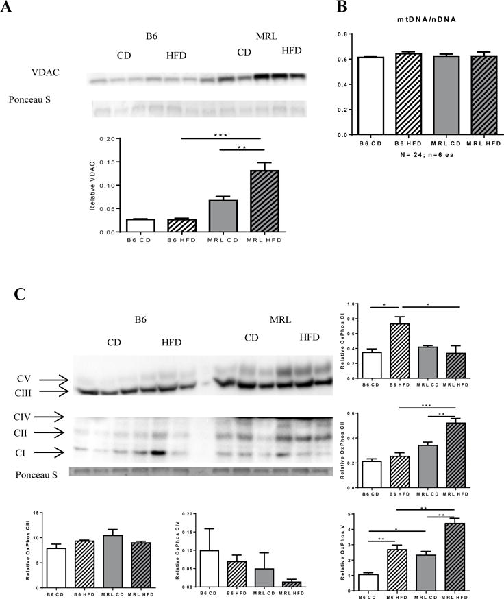
Figure 6.

MRL hepatocytes have increased mitochondrial proteins but not increased mitochondrial DNA. Liver lysates were western blotted and assessed for the mitochondrial protein Voltage-dependent anion channel (VDAC). (A) MRL mice have increased VDAC expression after HFD. (B) Quantification of mitochondrial DNA content over nuclear DNA through real time qPCR yielded no difference in mitochondrial content. (C) Further analysis on expression levels on oxidative phosphorylation chain proteins showed differential expression of proximal complexes I and II subunits between the two strains. Distal complexes III and IV subunits do not change in either strain, while nuclear encoded complex V subunit increases in both strains, with the MRL mice maintaining a higher basal expression. n=6−3; * p <0.05, ** p<,0.01 0.001 *** > p >0.0001.
