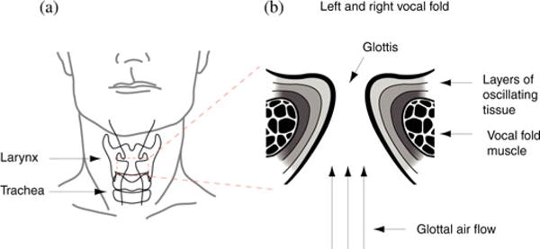
Fig. 1.

Vocal organ, the larynx, of humans. (a) The larynx is positioned in the upper neck area. It consists of a framework of cartilages, ligaments, and muscles. Inside the larynx are two vocal folds positioned. (b) Schematic of the enlarged left and right vocal folds. Active movements of larynx muscles narrow the space between the vocal folds (“glottis”) before voice is produced. Aerodynamic forces set vocal folds into self-sustained oscillations during expiration. The morphology of the vocal folds determines their oscillation behavior. Vocal folds in humans and many other mammals are multilayer structures. A deep layer (also called “body”) is the thyroarytenoid muscle. Three layers of connective tissue (“lamina propria”) and a layer of epithelium represent the tissue that is exposed to and interacting with the air flow (three arrows indicate air flow direction).
