Fig. 2.

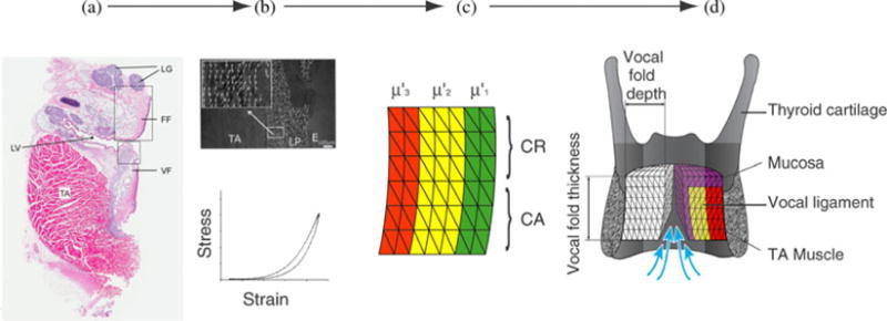
Compartmentalization of the vocal fold and implementation in a finite element model. (a) Differentially stained thin serial coronal sections are used to collect information about collagen and elastin content, fiber orientation, and hyaluronic acid content (e.g., [31], [35], [40], [41]). (b) The collagen fiber orientation is further investigated with greater detail using polarized light [36] (white arrows indicate collagen fibers). Mechanical tests are used to determine viscous and tensile strength of various compartments of the vocal fold [31], [35], [36]. (c) The vocal fold is discretized into compartments (superficial, intermediate, and deep, indicated here by three different colors; as well as upper, CR and lower, CA). A compartment is defined as a morphologically homogeneous portion of the vocal fold. It must not be smaller than what a surgeon can operate on (i.e., locate, manipulate, inject graft), but should be represented by enough finite elements to satisfy mesh requirements. Compartments are also created from anterior to posterior (not shown). Each compartment is characterized by geometry, fiber stress in the superficial , the intermediate , and deep layer .(d) Finally, the vocal fold structure is embedded into the laryngeal framework [13]. LV laryngeal ventricle; VF vocal fold; FF false fold; TA thyroarytenoid muscle; LG laryngeal glands; LP lamina propria; , , —shear modulus of respective layers.
