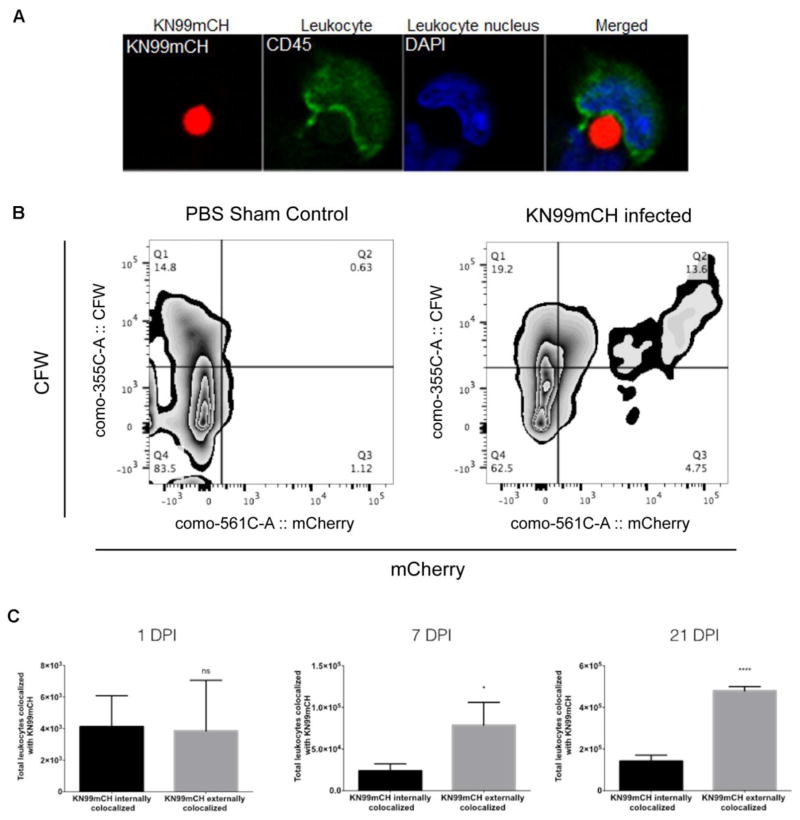
Fig. 8. KN99mCH fluorescence and cell staining for analysis of fungal: host interactions.
Mice were intranasally inoculated with either PBS (sham control) or 5 x 10⁴ KN99mCH and single-cell suspensions were prepared at on 1, 7, and 21 days PI from MDLN and lungs. (A) A KN99mCH cell being engulfed by a MDLN leukocyte (CD45-labeled) at day 21 PI. (B) Quantitation of externally or internally co-localized KN99mCH with host leukocytes and analysis strategy. Lung single-cell suspensions were stained with a fluorochrome-conjugated antibody towards CD45 (BioLegend), and were then stained with CFW (Sigma-Aldrich). Samples were then fixed (1% PFA), and then examined by flow cytometry. Following forward vs side scatter exclusion of cellular debris, Fluorescence minus one (FMO) controls were used to objectively set the gates to identify the leukocyte population (CD45+). This population was then further delineated for KN99mCH being internally (mCherry+CFW−) or externally (mCherry+CFW+) colocalized with leukocytes from 21 DPI. Rare cell events not shown. (C) To better understand the differences in colocalization of KN99mCH between those that were internally and externally colocalized with leukocytes, collected cells were compared on 1, 7, and 21 DPI. Statistical analysis was done using a two-tailed unpaired Student’s t test comparing KN99mCH externally colocalized to KN99mCH internally colocalized. Data is presented as the mean +/− standard deviation with an N = 3. *p < 0.05, and ****p < 0.0001.

