
Figure 3. Lactate-induced phenotypic modulation of hiPSC-vSMCs is mediated by monocarboxylic acid transporters (MCTs).
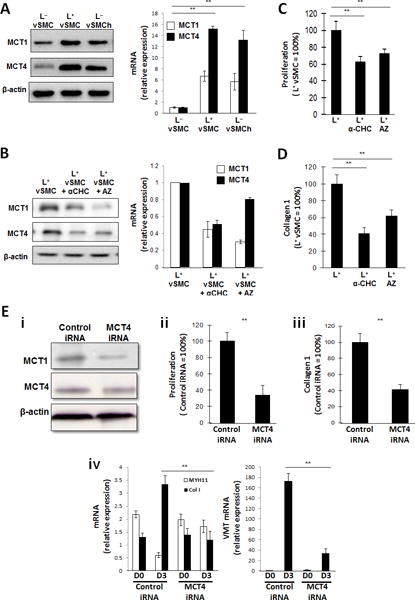
(A) hiPSC-derived vSMCs were cultured in L+ medium (L+vSMC), in L− medium (L−vSMC), or in L− medium under hypoxic conditions (L−vSMCh); then, the expression of MCT1 and MCT4 were evaluated via Western blot (left) and quantitative RT-PCR (right). mRNA levels were normalized to measurements in L−vSMC. (B-D) vSMCs were cultured in L+ medium, in L+ medium with the generalized MCT inhibitor α-CHC, or in L+ medium with the MCT1 inhibitor AZ3965 (AZ); then, (B) MCT1 and MCT4 expression was evaluated via Western blot (left) and quantitative RT-PCR (right), (C) proliferation was evaluated via proliferation assay (n= 3), and (D) collagen 1 levels were determined via ELISA (n= 3). mRNA levels were normalized to measurements in L+vSMC, and proliferation and collagen protein measurements were expressed as a percentage of measurements in cells that had been cultured in the absence of either inhibitor. (E) vSMCs were transfected with MCT4 iRNA or a scrambled iRNA sequence (Control iRNA) and cultured in L+ medium; then (i) MCT4 and MCT1 protein levels were evaluated via Western blot, (ii) proliferation was evaluated via proliferation assay (n= 3), and (iii) collagen 1 protein levels were evaluated via ELISA (n= 3); proliferation and collagen protein measurements were expressed as a percentage of measurements in cells that had been transfected with scrambled iRNA. (iv) MYH11, collagen 1 (Col 1), and vimentin (VMT) mRNA levels were evaluated via quantitative RT-PCR before (D0) and after (D3) a 3-day culture period (n= 3). **P<0.01 for all panels.