
Figure 2.
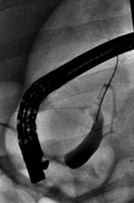
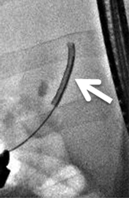
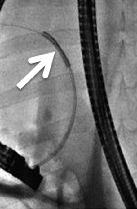
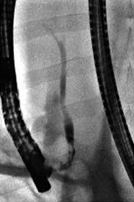
Intra-procedural cholangiography. A, Cholangiography was performed after cannulation of the guidewire into common bile duct. B, C, The IRE catheter was inserted into the common bile duct through the inner lumen of the endoscope. Arrows indicate the location of the electrode at the proximal and distal sites of treatment. D, The IRE catheter was removed after completion of pulse delivery. Retrograde cholangiography shows the immediate post-treatment patency of the bile duct and no evidence of extravasation of contrast material.