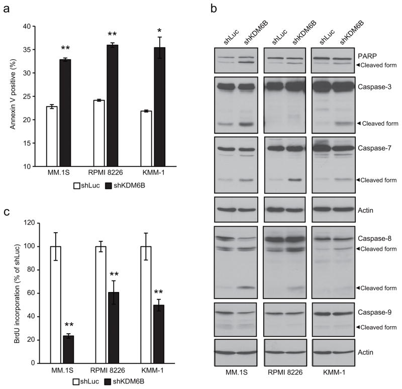
Figure 3.
Knockdown of KDM6B induces apoptosis and reduces cell proliferation in MM cells.
(a-c) MM.1S, RPMI 8226 and KMM-1 cells were transduced with shKDM6B or shLuc. (a) After 3 days (MM.1S) or 5 days (RRPMI 8226 and KMM-1) of infection, cells were analyzed for apoptosis by measuring annexin V-positive cells using the flow cytometer. Data represent mean ± s.d. of duplicate measurements. *P <0.05, **P < 0.01 compared with shLuc. (b) After 3 days (MM.1S) or 5 days (RRPMI 8226 and KMM-1) of infection, whole cell lysates were extracted and subjected to immunoblot analysis with the indicated antibodies. Actin served as the loading control for each membrane. (c) After 3 days of infection, the same number of viable cells were seeded and cultured for another 5 days. BrdU incorporation was then determined by BrdU assay. Data represent mean ± s.d. of triplicate cultures. **P < 0.01 compared with shLuc.

